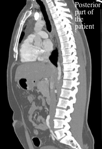

It also allows for unique imaging along an angled plane that can be used when requested by the referring physician or the radiologist. The CT gantry can be tipped cranially and caudally to deviate from the axial plane by 30 degrees in either direction and this can be used to correct for patient anatomic variability to maintain the axial plane, if necessary. These reformatted images are the result of a computer algorithm reformatting the original digital image data and, as such, these supplemental anatomic planes of imaging do not require additional x-ray exposure. Often the axial images obtained during the initial x-ray exposure will be digitally reformatted into other anatomic planes i.e. The standard orientation of image acquisition for CT is in the axial plane, hence the old name for this machine of CAT (computed axial tomography) scanner. The patient lies on the CT table in anatomic position and remains in that position throughout the study. The patient does not need to be moved, or re-positioned, to obtain CT images in different anatomic planes.

This imaging device is presented in Figure 3.21. This rotating x-ray source, associated with the rotating detectors, and the horizontally moving table top is called a helical CT scanner.

At the same time that the tube and detectors spin the horizontal table top that the patient lies on moves slowly through the gantry ring. The whole assembly that holds the x-ray tube and x-ray detectors is in the large doughnut shaped part of the unit called, the gantry. The detectors for the x-rays that pass through the patient are located in the ring structure and rotate in unison with the source of the x-rays. The x-ray tube generates x-rays only when programmed and activated by a CT Technologist. The x-ray tube is situated in a ring assembly that is able to spin around the horizontal table that the patient lies on. CT relies upon x-rays to facilitate image creation.


 0 kommentar(er)
